

An annular tear, also known as an annular fissure, represents a common spinal condition characterized by a tear in the annulus fibrosus—the tough, outer layer of an intervertebral disc. These tears often result from the natural wear and tear of aging or, less frequently, due to trauma or repetitive stress. Typically, an annular tear affects the lumbar region of the spine and can be a significant source of lower back pain and related symptoms. Understanding the anatomy, causes, symptoms, and management options for annular tears is essential for patients and healthcare providers alike.
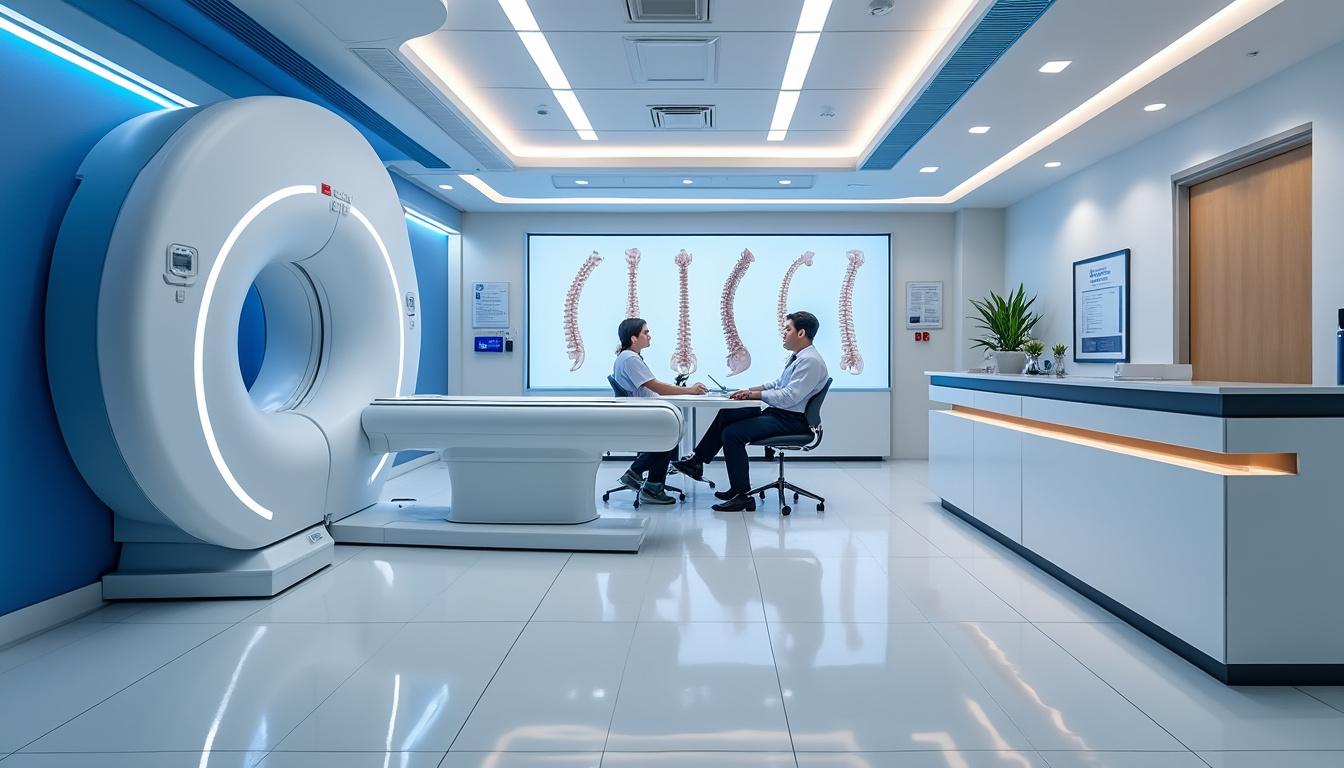
In recent years, advances in diagnostic imaging and minimally invasive treatments have broadened the scope of care for patients suffering from annular tears. Clinics like TAGMED in Québec emphasize a patient-centered approach, grounded in scientific rigor, innovation, and empathy, leveraging cutting-edge technologies such as spinal decompression therapy and regenerative medicine. This article explores annular tears in depth from the perspective of leading spine care experts, focusing on their causes, clinical presentations, evaluation methods, and the spectrum of available management strategies to help patients and practitioners make informed decisions.
Structural Insights into Annular Tears: Understanding the Intervertebral Disc and Annulus Fibrosus Function
The intervertebral disc serves a critical function in spinal health, located between vertebral bodies and acting as a shock absorber and stabilizer. An adult human spine comprises 23 intervertebral discs distributed along cervical, thoracic, and lumbar regions. Each disc is composed mainly of two elements: the nucleus pulposus and the annulus fibrosus. The nucleus pulposus is a gel-like core rich in water and proteoglycans, enabling it to absorb compressive forces effectively. Surrounding it is the annulus fibrosus, consisting of 15-20 concentric layers of type I collagen fibers configured in alternating oblique orientations that provide substantial tensile strength and protect the nucleus pulposus within.
One critical anatomical nuance is the posterior-lateral region of the annulus fibrosus, which contains vertically aligned collagen fibers. This region’s orientation creates a focal weakness, rendering it more susceptible to tears or fissures, especially under mechanical stress or degeneration. The intricate alternating patterns of the annulus layers ensure the disc can sustain multidirectional loads associated with bending, twisting, and compression. However, cumulative mechanical wear, biomechanical imbalance, or injury can cause fibers to separate or rupture, leading to an annular tear.
Key functions of the annulus fibrosus include:
- Containing the gel-like nucleus pulposus to maintain disc integrity
- Allowing flexibility and controlled movements of the spine such as flexion, extension, and rotation
- Protecting nerve roots by stabilizing disc pressure and preventing extrusion of disc material
| Intervertebral Disc Component | Structure | Function |
|---|---|---|
| Nucleus Pulposus | Gel-like core rich in water and proteoglycans | Absorbs compressive forces; maintains disc height |
| Annulus Fibrosus | 15-20 concentric collagen fiber layers; alternating oblique arrangement | Provides tensile strength; contains nucleus pulposus; stabilizes spine |
Understanding this anatomy is essential for interpreting how annular tears develop and their clinical implications. The tear compromises the containment function, which can then expose nociceptive nerve fibers in the annulus, causing pain and potentially leading to disc herniation if the nucleus pulposus extrudes through the defect.

Causes and Risk Factors Behind Annular Tears: From Age-Related Degeneration to Mechanical Stress
Annular tears primarily occur due to degenerative changes in the spine that accumulate with aging. Normal wear and tear subject the discs to repetitive loading, microtrauma, and biochemical shifts that progressively weaken the annulus fibrosus. Over time, dehydration of the nucleus pulposus reduces its cushioning ability while collagen fiber integrity in the annulus deteriorates, increasing susceptibility to fissures and tears.
While a single traumatic event may cause an annular tear, more often it is the result of chronic stress and compromised biomechanics. Posture defects, repetitive twisting or heavy lifting, smoking, and excess body weight are well-documented risk factors aggravating degeneration. Additionally, genetics may confer some predisposition to faster disc degeneration and thus earlier onset of annular tears.
Common causes and contributing factors include:
- Age-related biochemical and structural degeneration of disc tissue
- Chronic mechanical stress from work or sports activities
- Smoking, which impairs microcirculation to disc tissues
- Obesity increasing axial spinal load
- Genetic predisposition
In clinical practice, the Modified Dallas Classification is frequently employed to grade tear severity, based on imaging findings such as MRI or discography:
| Grade | Description of Annular Tear Severity |
|---|---|
| Grade 0 | No annular fissure detected |
| Grade 1 | Tear extends to the inner one-third of the annulus |
| Grade 2 | Tear reaches the middle one-third of the annulus |
| Grade 3 | Tear extends to the outer one-third of the annulus |
| Grade 4 | Tear with contrast tracking radially outward beyond annulus |
The anatomical susceptibility and the graded extent of tears guide both the prognosis and the management plans. Pathological tears (Grade 2 or higher) are more likely to cause symptoms or lead to complications such as disc herniation.
Clinical Manifestations and Symptoms of Annular Tears: Pain Patterns and Neurological Features
Annular tears exhibit a broad clinical spectrum, ranging from silent, incidental findings to severe, debilitating pain and neurological symptoms. Most annular fissures are asymptomatic, discovered incidentally during imaging performed for unrelated indications. However, when symptomatic, the condition can manifest as localized axial pain or radicular symptoms mimicking herniated disc presentations.
Localized symptoms generally arise from inflammatory reactions at the tear site, sensitizing nerve endings embedded within the annulus fibrosus. Patients often report deep, aching pain exacerbated by activities that increase spinal stress such as bending, twisting, coughing, or sneezing. Muscle stiffness or spasms and reduced spinal mobility frequently accompany the pain.
If the tear irritates or compresses adjacent spinal nerve roots, radicular symptoms develop, which may include radiating shooting pain, numbness, tingling, or muscle weakness following the nerve distribution. Symptoms usually correspond to the affected spinal level and can significantly impair quality of life.
Frequent symptoms associated with annular tears include:
- Lower back pain localized to the area over the affected disc
- Muscle stiffness and reflexive spasms
- Radiating pain down the leg or arm (radiculopathy) if nerve roots are involved
- Increased pain with spinal movements or increased intra-abdominal pressure (coughing, sneezing)
- Numbness, tingling, or weakness in extremities
Physical examination may reveal limited spinal range of motion or tenderness, but findings can be subtle if nerve root irritation is absent. The differentiation between simple annular fissure pain and accompanying nerve involvement is crucial as it impacts treatment strategies and prognosis.
| Symptom Type | Presentation | Clinical Correlation |
|---|---|---|
| Localized Pain | Deep, aching, increased by movement | Inflammation at annular tear site |
| Muscle Spasm | Reflexive tightening around lumbar spine | Protective response to pain |
| Radicular Pain | Shooting, sharp, follows nerve roots | Nerve root irritation or compression |
| Neurological Deficits | Numbness, weakness, altered reflexes | Severe nerve root or cord involvement |
Clinical Case Example
A 45-year-old office worker experienced gradually worsening lower back pain over several months, exacerbated by prolonged sitting and forward bending. MRI revealed a Grade 3 posterior-lateral annular tear at L4-L5 without disc herniation. Initial management focused on conservative measures that successfully reduced inflammation and improved function. This example underscores the importance of accurate imaging and comprehensive clinical correlation.
Diagnostic Procedures for Annular Tears: Imaging Modalities and Clinical Evaluation
Correctly diagnosing an annular tear requires a combination of thorough patient history, physical examination, and advanced imaging techniques. Magnetic resonance imaging (MRI) remains the gold standard for identifying annular fissures due to its superior soft tissue contrast and ability to visualize disc structures, including the characteristic high-intensity zones (HIZ) associated with tears.
On T2-weighted MRI scans, annular tears appear as hyperintense (bright) lines within the normally hypointense (dark) annulus fibrosus, reflecting increased water content or inflammation at the fissure. The Modified Dallas Classification applies to MRI and discography to classify tear severity, aiding in therapeutic decision-making.
Computed tomography (CT) scans provide excellent bony detail but are less sensitive for annular fissures. However, CT myelography can be valuable in demonstrating nerve root compression when MRI is contraindicated or inconclusive.
While discography—injecting contrast into the disc—was once widely used to identify painful discs by reproducing concordant pain, it has fallen out of favor due to high false-positive rates and potential to accelerate disc degeneration. Its current use is reserved for complex preoperative planning when MRI findings are ambiguous.
Diagnostic tools and their utility include:
- MRI T2-weighted sequences: primary for visualizing annular tears and disc pathology
- CT myelogram: alternative for nerve root compression visualization
- Plain radiographs: limited usefulness for soft tissue but assess disc space and spinal alignment
- History and physical exam: critical for correlating symptoms with imaging
- Discography: rarely used; reserved for select cases with unclear diagnosis
| Diagnostic Method | Advantages | Limitations |
|---|---|---|
| MRI | Non-invasive, detailed soft tissue visualization, detects HIZ | Cost, contraindications (pacemakers, metal implants) |
| CT Myelogram | Good for nerve root visualization if MRI unavailable | Invasive, radiation exposure |
| Discography | Functional assessment by reproducing pain | High false-positive rate, invasive |
| Physical Examination | Non-invasive, guides suspicion | Non-specific findings |
Conservative Treatment Strategies for Annular Tears: Prioritizing Safety and Efficacy
Given the often-benign natural history of annular tears, conservative treatment remains the cornerstone for patients without significant neurological deficits or severe pain. This approach emphasizes reducing inflammation, alleviating symptoms, and enhancing spinal function through non-invasive measures.
Non-steroidal anti-inflammatory drugs (NSAIDs) are widely used to mitigate inflammatory responses at the tear site, providing pain relief and improved mobility. Additionally, physical therapy focusing on low-impact exercises and core stabilization plays an essential role in promoting healing and preventing recurrence. These exercises strengthen paraspinal muscles, improve posture, and enhance spinal biomechanics to reduce undue stress on the annulus fibrosus.
Additional conservative modalities include activity modification, ergonomic interventions, and patient education on lifestyle adaptations to reduce mechanical strain on the spine. Weight management and smoking cessation further support disc health and healing.
Common conservative treatment components include:
- NSAIDs for pain and inflammation control
- Physical therapy emphasizing core strengthening and flexibility
- Ergonomic adjustments at work and home
- Activity modification to avoid aggravating movements
- Lifestyle changes such as weight loss and smoking cessation
| Conservative Therapy | Purpose | Expected Outcome |
|---|---|---|
| NSAIDs | Reduce inflammation and pain | Symptom relief, improved mobility |
| Physical Therapy | Strengthen muscles, improve stability | Prevention of recurrence, functional improvement |
| Activity Modification | Avoid exacerbating actions | Reduced symptom flare-ups |
| Lifestyle Changes | Enhance disc health | Long-term spinal resilience |
Patient adherence to these conservative measures is crucial. Dr. Sylvain Desforges stresses personalized evaluation and targeted interventions to optimize outcomes, aligning with rigorous scientific standards and ethical patient care.
Advanced Therapeutic Techniques for Annular Tears: Minimally Invasive and Regenerative Approaches
In cases where conservative management fails, and symptoms persist or progress, several minimally invasive procedures have emerged as effective options. These techniques aim to directly address annular fissures by reducing inflammation, denervating pain fibers, and promoting tissue healing, while avoiding open surgery’s risks.
Intradiscal electrothermal therapy (IDET) uses controlled heat delivered via a catheter to denature nociceptive nerve fibers and cause collagen shrinkage in the annulus fibrosus. Although promising initially, IDET has produced variable results, limiting widespread acceptance. Alternatively, biacuplasty utilizes bipolar radiofrequency ablation, offering more precise thermal therapy to the posterior annulus, with clinical studies demonstrating meaningful pain reduction in select discogenic back pain patients.
The Disc-FX procedure exemplifies an innovative, minimally invasive intervention combining nucleus ablation, annuloplasty, and nucleus modulation using radiofrequency energy. Performed under fluoroscopic guidance through Kambin’s triangle access, Disc-FX targets pain-generating sites while preserving disc integrity, providing a bridge between conservative care and open surgery.
Advanced treatments available include:
- Intradiscal Electrothermal Therapy (IDET)
- Biacuplasty (radiofrequency ablation)
- Disc-FX procedure involving radiofrequency nucleus modulation
- Local steroid injections for inflammation control
| Procedure | Mechanism | Indications | Limitations |
|---|---|---|---|
| IDET | Heat denatures nerve fibers, shrinks collagen | Symptomatic annular tears without large herniations | Mixed evidence, variable efficacy |
| Biacuplasty | Bipolar radiofrequency ablation | Discogenic low back pain, annular fissures | Requires precise patient selection |
| Disc-FX | Radiofrequency nucleus ablation and annuloplasty | Contained herniations, discogenic pain | Limited long-term data |
| Steroid Injections | Anti-inflammatory effect | Inflammatory symptoms | Temporary relief, potential adverse effects |

Regenerative Medicine and Emerging Therapies: The Future of Annular Tear Management
Regenerative medicine offers promising avenues for annular tear treatment by harnessing biological agents to stimulate healing, reduce inflammation, and perhaps restore disc structure. Among the most studied modalities are platelet-rich plasma (PRP) and stem cell therapies.
PRP injections utilize concentrated autologous platelets releasing growth factors and anti-inflammatory cytokines, potentially enhancing tissue repair within the damaged annulus fibrosus. Early clinical trials demonstrate significant pain reduction and functional improvement over 6 to 12 months.
Stem cell-based therapies, particularly those utilizing bone marrow-derived mesenchymal stem cells (BMSCs), show potential for disc matrix regeneration. These cells may differentiate into fibrocartilage-producing cells, promoting structural repair. Although still investigational and not widely covered by insurers, emerging evidence from institutions such as Mayo Clinic and Cleveland Clinic supports their cautious application in carefully selected patients.
Highlights of regenerative treatments for annular tears:
- Platelet-Rich Plasma (PRP) injections targeting inflammation and healing
- Mesenchymal stem cell therapies encouraging matrix regeneration
- Complementary to conventional therapies, not standalone cures
- Currently considered investigational with ongoing research
| Regenerative Therapy | Mechanism | Evidence Status | Clinical Status |
|---|---|---|---|
| Platelet-Rich Plasma (PRP) | Growth factors reduce inflammation, promote repair | Preliminary positive results; limited large-scale studies | Off-label use; not FDA-approved for intradiscal injection |
| Stem Cell Therapy | Cell differentiation, matrix regeneration | Experimental; early-phase clinical trials promising | Investigational; limited availability |
Prognosis and Long-Term Outlook: Navigating Recovery and Prevention
The course of annular tears is generally favorable, especially when managed early and conservatively. Many individuals experience symptomatic relief as inflammation resolves and muscular strength improves. Nevertheless, the presence of an annular tear increases the disc’s vulnerability to future injury and degeneration, requiring ongoing attention to spinal health.
Successful rehabilitation involves not only symptom control but also prevention strategies to minimize re-injury risks. Patients should be educated on proper posture, lifting techniques, and core stabilization exercises tailored to their daily activities. Routine follow-up and multidisciplinary care at facilities embracing evidence-based protocols, such as those led by Dr. Sylvain Desforges, provide patients a pathway to durable spine health.
Key factors influencing prognosis include:
- Severity and location of the annular tear
- Presence of associated disc herniation or nerve root involvement
- Promptness and appropriateness of treatment
- Patient adherence to rehabilitation and lifestyle modifications
- Overall health and comorbidities influencing tissue healing
| Prognostic Factor | Impact on Recovery |
|---|---|
| Mild annular fissure | Often spontaneous improvement |
| Severe tear with herniation | May require surgical intervention |
| Early intervention | Better symptom control, functional recovery |
| Adherence to physical therapy | Reduced recurrence, improved stability |
| Presence of comorbidities | Potential delayed healing |
Comprehensive Patient Support and Expert Guidance in Spine Care
At the forefront of spinal care innovation and compassionate patient management, Dr. Sylvain Desforges exemplifies an integrated approach combining expertise, scientific rigor, and empathy. Drawing upon over 30 years of clinical experience and leadership roles in key institutions such as the Canadian College of Osteopaths and Clinique TAGMED, Dr. Desforges tailors comprehensive evaluations that respect each patient’s unique situation.
His protocols utilize advanced diagnostic modalities aligned with standards from the Collège des médecins du Québec and incorporate emerging technologies, including neurovertebral decompression and novel implant systems by leaders like Medtronic, Stryker, NuVasive, Zimmer Biomet, and Boston Scientific. The goal is to ensure safety, efficacy, and transparent communication to empower patients and optimize outcomes.
Personalized care plans acknowledge the varying presentations and complexities of annular tears, assuring patients receive clear information regarding expectations, treatment options, and follow-up. This blend of innovation with ethical practice fosters confidence and facilitates recovery journeys well beyond initial symptom relief.
Core principles emphasized by expert spinal care providers include:
- Patient-centered evaluation and individualization of treatment
- Adherence to evidence-based guidelines and regulatory frameworks
- Education focusing on clear, accessible explanation of pathology and treatments
- Integration of cutting-edge technologies with traditional osteopathic care
- Empathetic communication fostering motivation and trust
| Principle | Implementation in Clinical Practice |
|---|---|
| Scientific Rigor | Use of evidence-based protocols and continuous research |
| Ethical Compliance | Alignment with Canadian medical regulatory standards |
| Patient Education | Accessible explanations, personalized consultations |
| Innovative Technology | Advanced diagnostics and minimally invasive therapies |
| Compassionate Care | Listening, empathetic interaction, motivational support |
What symptoms differentiate an annular tear from a herniated disc?
While both conditions can cause similar symptoms such as localized pain and radiculopathy, an annular tear refers to a tear in the disc’s outer layer and may occur without herniation. Herniated discs involve the nucleus pulposus protruding beyond the annulus and often result in more pronounced nerve compression symptoms. Definitive differentiation requires imaging studies like MRI.
Can an annular tear heal on its own?
Mild annular tears can be asymptomatic and may heal or stabilize over time with conservative management such as physical therapy and anti-inflammatory medications. However, severe tears, especially those accompanied by disc herniation, often require more advanced treatments.
What are the risks of surgical treatment for annular tears?
Surgical interventions carry risks such as infection, nerve damage, and recurrence of symptoms. Surgery is generally reserved for cases with severe pain, neurological deficits, or failed conservative treatment. Minimally invasive procedures offer alternative options with reduced recovery times.
How does regenerative medicine work in treating annular tears?
Regenerative treatments like PRP and stem cell therapy aim to reduce inflammation and promote tissue repair at the annular tear site. These therapies are investigational and currently supported by emerging clinical evidence. They are used alongside conventional treatments rather than as standalone solutions.
When should I seek medical evaluation for suspected annular tear?
If you experience persistent back pain lasting more than a few weeks, radiating limb symptoms, or neurological changes such as numbness or weakness, it is important to consult a healthcare provider promptly for comprehensive evaluation and management tailored to your condition.