

Low back pain is one of the most common and challenging health complaints globally, affecting millions of individuals each year. Its causes range from simple muscle strain to complex spinal disorders, demanding a nuanced and thorough diagnostic approach. Navigating this terrain requires expert evaluation, integrating diverse medical perspectives and advanced diagnostic technologies to pinpoint the underlying causes precisely. Such a comprehensive process ensures tailored treatment plans that optimize recovery and minimize unnecessary interventions.
In the realm of spinal care, experts like Dr. Sylvain Desforges play a crucial role, combining over three decades of clinical experience with scientific rigor and compassionate patient engagement. His dedication to evidence-based practice and innovative therapeutic methods, including advanced spinal decompression techniques, sets a benchmark for diagnosing and managing low back pain effectively. By demystifying the evaluation pathways and leveraging the latest in imaging and neurological assessment, Dr. Desforges guides patients through their journey with clarity and personalized attention.
Comprehensive Medical History and Symptom Review for Low Back Pain Diagnosis
A fundamental step in diagnosing low back pain involves a detailed review of the patient’s medical history and a nuanced understanding of their symptoms. This foundational phase, often conducted as an in-depth conversation, reveals valuable clues about the pain’s onset, characteristics, and impact on daily living.
During this consultation, the clinician explores several dimensions of the patient’s experience:
- Pain characteristics: When did the pain begin? Was it sudden or gradual? What activities or postures affect its intensity?
- Duration and patterns: Is the pain acute, chronic, or recurrent? Identifying these timelines helps distinguish between transient issues and chronic conditions.
- Location and radiation: The precise area of discomfort is noted along with any radiation to limbs, which may indicate nerve involvement such as sciatica.
- Quality and intensity: Patients describe their pain using terms like sharp, dull, burning, or stabbing, revealing possible tissue types involved.
- Physical activity and lifestyle assessment: Understanding occupational demands, exercise habits, and sedentary time offers insight into contributing factors.
- Sleep and psychosocial elements: Sleep quality and any presence of anxiety or depression are documented, recognizing their influence on pain perception.
- Previous treatments and outcomes: Past interventions and their success or failure can guide current management decisions.
- Red flags for serious conditions: Specific symptoms like unexplained weight loss, fever, or neurological impairments necessitate further investigation.
This intricate puzzle assembled from patient history demands an expert’s keen eye to discern subtle patterns or warning signs. Dr. Sylvain Desforges emphasizes the importance of these initial findings to avoid unnecessary testing and to tailor individualized care effectively.
| Aspect of History | Purpose | Example Questions |
|---|---|---|
| Onset and Duration | Define if pain is acute or chronic | “When did the pain start?” “Has it been continuous or intermittent?” |
| Pain Characteristics | Identify involved tissues or nerves | “Is the pain sharp, burning, or dull?” |
| Aggravating/Relieving Factors | Determine triggers and effective reliefs | “Does sitting worsen the pain?” “Is heat or ice helpful?” |
| Associated Symptoms | Identify red flags or neurological involvement | “Have you experienced numbness or bladder issues?” |
| Lifestyle & Occupation | Contextualize risk and causative factors | “Do you have a physically demanding job?” |

Hands-On Physical Examination: Key Steps and Techniques in Evaluating Low Back Pain
Once a comprehensive medical history is established, the physical examination serves as the clinician’s tactile and observational toolkit to uncover crucial signs that symptoms alone cannot reveal. This stage involves careful assessment of posture, spinal mobility, muscle condition, and neurological function.
The physical exam typically encompasses the following components:
- Observation of posture and spinal curvature: Identifying deviations such as scoliosis, kyphosis, or altered lordosis that may influence pain patterns.
- Palpation: Gentle manual inspection of muscle tenderness, spasm, or skeletal abnormalities along the lumbar spine.
- Range of motion tests: Assessing flexibility and detecting movements that exacerbate pain, including forward flexion, extension, lateral bending, and rotation.
- Neurological assessment: Testing motor strength, sensation, and reflexes in lower limbs to detect nerve root irritation or compression.
- Provocative maneuvers: The straight leg raise test, for example, can implicate sciatic nerve irritation due to herniated discs.
For neurological testing, Dr. Desforges emphasizes examining key nerve roots (L4, L5, S1), which are often the culprits in lower back pain radiating to the legs. Careful evaluation allows early detection of red flags like progressive motor weakness or saddle anesthesia, which require urgent medical attention.
| Physical Test | Purpose | Clinical Significance |
|---|---|---|
| Posture and Curvature Observation | Detect spinal deformities | May indicate scoliosis or compensatory postural adaptations |
| Palpation of Spine/Muscles | Identify tenderness or spasm | Indicates muscle strain or vertebral issues |
| Range of Motion Tests | Determine flexibility and pain triggers | Limitations suggest mechanical or neurological causes |
| Neurological Testing | Assess nerve function | Detects nerve root compression or neuropathy |
| Straight Leg Raise Test | Elicit sciatic nerve irritation | Positive result suggests lumbar herniated disc |
The detailed hands-on examination enables experts like Dr. Desforges to connect historical clues with physiological signs, ensuring a targeted and personalized diagnostic process that precedes any imaging.
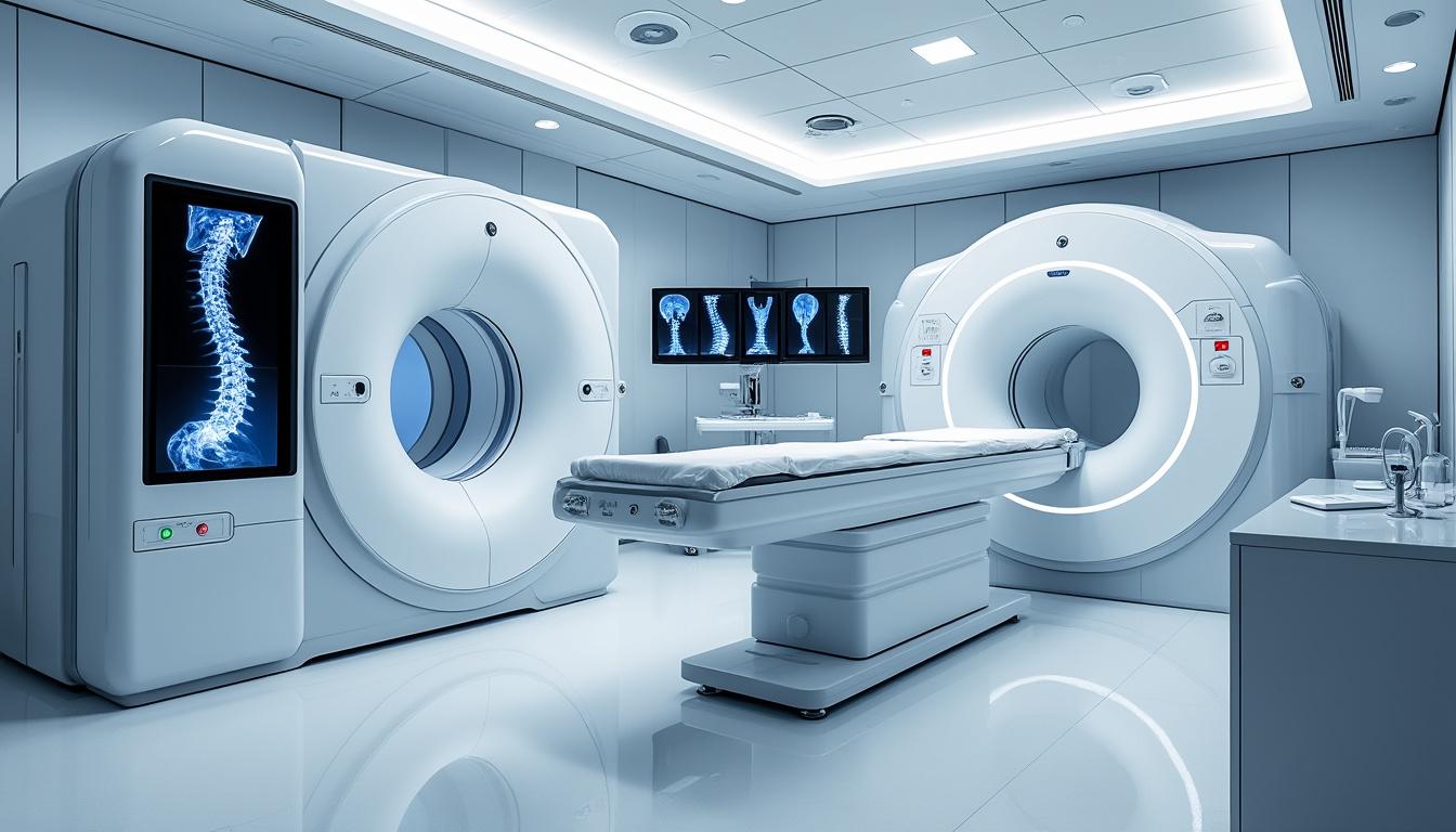
The Critical Role of Imaging in Low Back Pain Diagnosis: When and Why?
In the evaluation of low back pain, the judicious use of imaging technologies is paramount. While many cases of acute low back pain resolve spontaneously without the need for imaging, persistent or complicated symptoms often require advanced visualization for accurate diagnosis and treatment planning.
Common imaging modalities used include:
- X-rays: Useful for identifying bone abnormalities like fractures, arthritis, or scoliosis.
- MRI (Magnetic Resonance Imaging): Provides detailed images of soft tissues including intervertebral discs, ligaments, and nerves without radiation exposure.
- CT scans: Offers cross-sectional views valuable for complex bone or joint disorders.
- Myelography: Combined with CT, helps visualize spinal canal and nerve root impingement.
Clinical guidelines from organizations like the American College of Physicians recommend avoiding routine imaging for non-specific acute low back pain, emphasizing its use when red flags or failed conservative treatments are present. This approach reduces unnecessary exposure, overdiagnosis, and healthcare costs.
Dr. Desforges advocates for employing state-of-the-art equipment from leading healthcare technology manufacturers, including Siemens Healthineers and Philips Healthcare, ensuring the highest quality diagnostic imagery. He closely monitors evolving evidence, emphasizing imaging only when it meaningfully impacts clinical decisions.
| Imaging Modality | Primary Diagnostic Use | Advantages | Limitations |
|---|---|---|---|
| X-ray | Bone structure and alignment | Widely available, low cost | Poor soft tissue detail, radiation exposure |
| MRI | Soft tissue and neural structures | No radiation, highly detailed images | Costly, limited availability, contraindications (e.g., pacemakers) |
| CT Scan | Bone and complex anatomy | Detailed, rapid imaging | Higher radiation, less soft tissue detail than MRI |
| Myelography | Spinal canal and nerve roots | Effective for nerve blockage detection | Invasive, requires contrast injection |
Red Flags and Symptoms Indicating Serious Back Conditions
While the majority of low back pain cases are benign and self-limiting, identifying symptoms that warning of potentially serious underlying disorders is crucial. These “red flags” signal the need for urgent diagnostics and possibly immediate intervention.
Common red flags include:
- Age extremes: Onset of pain before age 20 or after age 55 may indicate atypical causes.
- History of cancer: Raises suspicion for metastatic disease.
- Unexplained weight loss or fever: Potentially indicative of infection or malignancy.
- Trauma: Significant injury preceding onset may lead to fracture.
- Neurological deficits: Including numbness in the saddle area, bladder or bowel dysfunction, and progressive leg weakness.
- Immunosuppression or intravenous drug use: Heightened infection risk.
A rapid and robust evaluation protocol led by experts such as Dr. Desforges prioritizes these signs to prevent delays in treatment of conditions like cauda equina syndrome, spinal infections, or neoplasms.
| Red Flag | Implications | Clinical Action |
|---|---|---|
| Bladder or bowel dysfunction | Cauda equina syndrome | Immediate emergency referral |
| Unexplained weight loss | Possible malignancy | Urgent imaging and oncology consult |
| Fever with back pain | Spinal infection | Blood tests, imaging, antibiotics |
| History of trauma | Vertebral fracture | Imaging and orthopedic referral |
By maintaining vigilance for these warning signs and integrating patient history with exam findings, Dr. Desforges ensures patient safety while guiding appropriate investigations and therapeutic decisions.
Advanced Neurodiagnostic Testing in Assessing Low Back Pain
When clinical examination and imaging leave questions unanswered, additional neurodiagnostic tests offer valuable insights into nerve function and pain origin. These tests include electrodiagnostic studies such as electromyography (EMG) and nerve conduction studies (NCS).
Such assessments help:
- Identify nerve root irritation or damage.
- Differentiate between peripheral neuropathy and radiculopathy.
- Localize the level of nerve involvement to refine treatment plans.
For example, if a patient exhibits leg weakness and numbness but imaging shows equivocal disc abnormalities, these tests can determine whether the nerve root is truly compromised. Dr. Desforges integrates these investigations thoughtfully, balancing the benefits with patient comfort and clinical necessity.
| Test | Purpose | Typical Findings |
|---|---|---|
| Electromyography (EMG) | Evaluate muscle electrical activity | Signs of denervation or chronic nerve damage |
| Nerve Conduction Study (NCS) | Measure nerve signal speed | Delay or block in nerve conduction |
| Somatosensory Evoked Potentials (SSEP) | Assess sensory pathway integrity | Altered potential amplitudes indicating dysfunction |
The Integral Role of Specialist Evaluation without Medical Diagnosis
Dr. Sylvain Desforges embodies the essential role of an expert in spinal care who conducts comprehensive evaluations rather than offering formal medical diagnoses. His meticulous assessments aim to clarify the complex interplay of spinal structures and guide patients toward optimal non-surgical treatment pathways.
His approach includes:
- Detailed clinical interview and history taking emphasizing patient uniqueness.
- Comprehensive physical examination integrating osteopathic principles.
- Collaboration with diagnostic imaging centers utilizing equipment from Siemens Healthineers and Canon Medical Systems to ensure high diagnostic fidelity.
- Evidence-based recommendations tailored to patient circumstances.
- Patient education encouraging understanding and active participation in care.
This model prioritizes safety, ethics, and innovation. Patients benefit from a personalized care pathway founded on a thorough and scientifically rigorous evaluation, avoiding premature reliance on surgery or unnecessary imaging procedures.

Case Study: Non-Surgical Spinal Decompression Therapy and Low Back Pain
Breakthroughs in non-invasive spinal treatments such as spinal decompression therapy have transformed management options for patients with herniated discs and chronic low back pain. A prospective randomized controlled study documented on sosherniateddisc.com highlights significant pain reduction and functional improvement following this therapy.
Spinal decompression applies controlled mechanical traction, gently stretching the spine to relieve pressure on compressed nerves and discs. Dr. Desforges integrates this therapy at Clinique TAGMED, leveraging state-of-the-art equipment to customize treatment sessions based on precise patient evaluation.
Benefits of this approach include:
- Reduction in pain without the need for surgery or medications with heavy side effects.
- Improved spinal mobility and posture correction.
- Enhanced patient quality of life through targeted and patient-centric interventions.
- Aiding in long-term prevention of flare-ups, supported by patient education.
Patients report not only symptomatic relief but an empowering sense of reclaiming control over their health, underpinning the importance of innovation combined with tailored expertise.
Guiding Patients Through Diagnostic Complexities: The Importance of Clear Communication
Clear and accessible education lies at the heart of Dr. Desforges’ philosophy. Patients receive carefully explained information about their diagnostic findings, treatment options, and anticipated outcomes, empowering them to make informed decisions.
Key communication strategies include:
- Plain language explanations: Avoiding jargon, clarifying terms like “herniated disc” or “nerve root compression”.
- Discussion of benefits and limitations of diagnostics: Explaining why imaging may or may not be necessary.
- Emphasizing personalized care: Recognizing that each patient’s condition is unique and requires individualized evaluation.
- Patient reassurance and motivation: Encouraging questions and highlighting potential for recovery.
- Facilitating access to trusted resources: such as information on low back pain diagnosis and conservative treatment pathways on reputable sites.
This approach fosters trust and collaboration, essential elements for effective long-term management of low back pain.
| Communication Aspect | Purpose | Example |
|---|---|---|
| Plain Language | Make complex concepts accessible | “Your disc is like a cushion between vertebrae that can sometimes slip out of place.” |
| Benefit-Limitation Discussion | Set realistic expectations | “X-rays won’t show nerve issues, but MRI can.” |
| Personalization | Address individual needs | “Your pain pattern suggests a different approach than someone else’s.” |
| Encouragement | Build patient confidence | “Many patients improve with conservative care without surgery.” |
Partnering with Advanced Technology Companies to Improve Spinal Diagnostics and Care
The integration of leading medical technology firms into spinal care operations plays a critical role in enhancing diagnosis and treatment. Collaborations with industry leaders such as GE Healthcare, Fujifilm Healthcare, Hologic, BD (Becton, Dickinson and Company), and Zimmer Biomet enable continuous innovation and access to cutting-edge diagnostic tools and therapies.
Examples of advanced technologies improving patient outcomes include:
- Dynamic spinal implants: Supporting mobility while stabilizing the spine after injury or degeneration.
- Laser therapy modalities: Promoting tissue healing and pain relief through non-invasive procedures.
- Neurovertebral decompression technology: Allowing precision targeting of affected spinal segments.
- Advanced digital imaging: Facilitating higher-resolution views of spinal anatomy for accurate diagnosis.
- Data integration software: Enhancing clinical decision-making and personalized treatment planning.
These advancements underscore the commitment of experts like Dr. Desforges and the organizations he leads, such as the Canadian College of Osteopaths and the Alliance Canadienne de Médecine Alternative, to perpetual improvement in spinal care delivery.
What are the primary steps in diagnosing low back pain?
Diagnosis typically starts with detailed medical history and symptom review, followed by a physical examination. Imaging and further tests are used if necessary to identify serious conditions or persistent pain.
When is imaging recommended during low back pain evaluation?
Imaging is recommended when red flags are present, symptoms persist beyond a few weeks despite treatment, or to plan interventional procedures. Routine imaging for acute, non-specific pain is generally avoided.
What are ‘red flags’ in low back pain diagnosis?
Red flags are warning signs such as unexplained weight loss, fever, neurological deficits, or trauma history that indicate potentially serious underlying conditions requiring urgent assessment.
How does spinal decompression therapy help low back pain?
Spinal decompression therapy gently stretches the spine to relieve pressure on discs and nerves, reducing pain and improving mobility without surgical intervention.
Why is personalized evaluation important in managing low back pain?
Because low back pain causes vary greatly among individuals, a tailored evaluation helps identify the exact cause and select the most effective treatment, improving outcomes and patient satisfaction.