

Disc Herniation in Mirabel: Comprehensive Program Duration and Neurovertebral Decompression Therapy
June 13, 2026
Hospitals and Surgeon Collaborations: Insights and Experiences Shared
June 13, 2026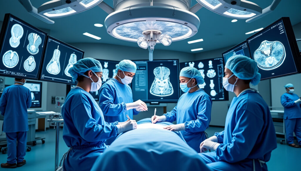
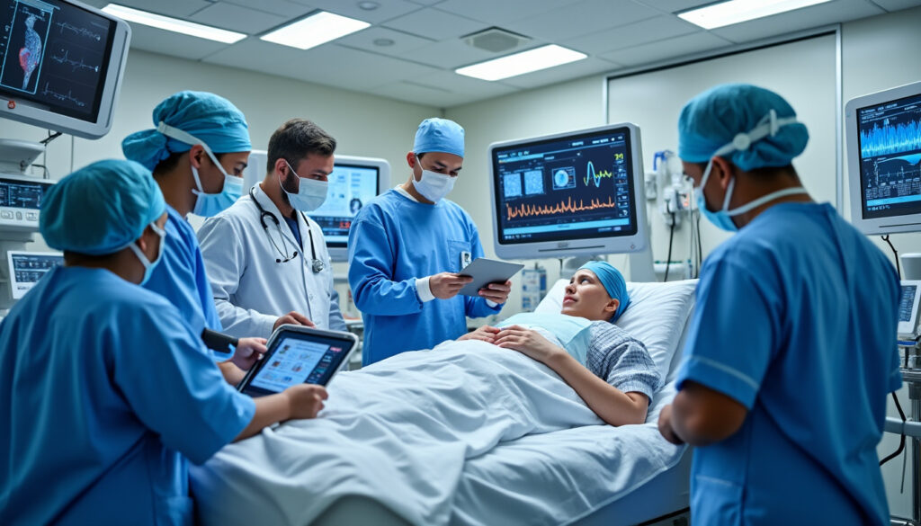
The landscape of surgery is undergoing a transformative evolution, driven significantly by the strategic management of medical imaging exams. As healthcare technology advances, the integration and organization of imaging diagnostics emerge as crucial pillars supporting surgical innovation and enhancing patient outcomes. This paradigm shift is most evident in complex surgical fields, such as spinal surgery, where precise anatomical visualization informs clinical decision making and surgical planning to an unprecedented degree. The ability to harness imaging technologies effectively not only streamlines preoperative preparation but also enriches intraoperative guidance and postoperative monitoring, setting new benchmarks for safety and efficacy.
In 2026, forward-thinking medical institutions increasingly recognize that the future of surgery is tightly intertwined with optimizing imaging protocols and coordination. This shift transcends the mere acquisition of images to encompass the orchestration of imaging modalities that provide real-time insights and foster collaborative communication among multidisciplinary teams. By capitalizing on innovations such as 3D functional MRI, computer-assisted navigation, and artificial intelligence, surgeons navigate the fine line between invasive procedures and minimally traumatic interventions, ensuring superior clinical results. These developments are bolstered by international collaborations that facilitate access to cutting-edge techniques and disseminate best practices in imaging management, elevating healthcare standards worldwide.
How Strategic Management of Imaging Exams Revolutionizes Surgical Planning
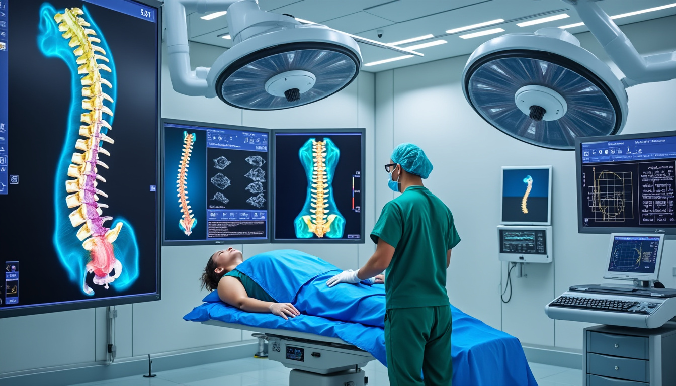
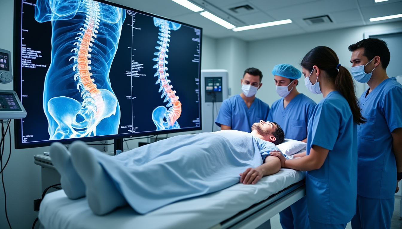
Effective surgical planning hinges on the availability and quality of medical imaging. Strategic management of imaging exams ensures that clinicians have timely access to high-resolution, clinically relevant data tailored to the specific needs of each patient. In spinal surgery, for example, preoperative imaging—ranging from conventional MRI and CT scans to advanced 3D functional MRI—enables detailed mapping of neural structures, lesion extents, and biomechanical dynamics. These insights inform the choice between surgical approaches such as microdiscectomy, spinal fusion, or motion-preserving prosthetics, optimizing risk-benefit profiles.
An organized imaging workflow reduces delays in decision making by harmonizing protocols between referring centers and surgical units. For patients traveling internationally for specialized care, as seen in collaborations between Quebec and French centers, the coherent transfer and evaluation of imaging studies are vital. Coordination experts meticulously combine existing images with additional functional exams, such as neurofeedback-assisted biofeedback, producing personalized profiles that guide surgeons with precision.
Moreover, aligning imaging diagnostics with surgical innovation accelerates the adoption of image-guided techniques. For instance, computer-assisted navigation leverages real-time imaging inputs to permit millimeter-accurate intervention while minimizing exposure and incision size. This approach depends on integrating intraoperative imaging platforms seamlessly into surgical workflows, a feat achievable only through robust exam management systems.
The interplay between preoperative imaging and clinical decision making exemplifies how strategic imaging exam management elevates patient outcomes. Organized imaging protocols reduce unnecessary repetition, focus diagnostic efforts on clinically meaningful targets, and enhance multidisciplinary communications. This not only augments surgical accuracy but also instills greater patient confidence through transparent, data-driven planning.

Understanding Sciatic Pain: Key Symptoms and When to Seek Medical Advice
Sciatic pain is a prevalent condition impacting a wide demographic, manifesting primarily as intense pain radiating from the lower back down the leg. This pain stems from irritation or compression of the sciatic nerve, a major nerve extending through the…
Integrating Imaging Diagnostics with Surgical Innovation for Enhanced Patient Outcomes
Medical imaging stands at the forefront of surgical innovation, showcasing the potential to transform patient outcomes through precision and safety. Advanced modalities like functional MRI, fluorescence-guided imaging, and robotic-assisted navigation have transcended traditional boundaries, embedding themselves into intricate surgical procedures. This integration renders visible real-time dynamics otherwise imperceptible, fostering minimally invasive techniques with superior preservation of neurological function.
The evolution from static, preoperative images to dynamic, intraoperative imaging exemplifies this trend. Surgeons now utilize augmented reality overlays and extended reality platforms to interpret medical imaging intuitively in three dimensions, enhancing spatial understanding and procedural accuracy. Such innovations are documented to reduce complication rates and accelerate recovery.
For example, spinal interventions increasingly use near-infrared fluorescence to map lymphatic structures accurately, a critical factor in oncological surgeries where tissue preservation impacts prognosis. The coupling of imaging diagnostics with robotic systems, such as the Da Vinci platform or the eCential 3D navigation robot, exemplifies the future of surgery, where precise, computer-mediated maneuvers reduce surgeon fatigue and enhance dexterity in confined anatomical spaces.
Strategic management of imaging exams supports these innovations by ensuring data fidelity and readiness. Centralized imaging databases and artificial intelligence facilitate image interpretation and predictive modeling, assisting surgeons and healthcare teams in adapting surgical strategies dynamically. This systemic integration bolsters patient outcomes by reducing intraoperative errors and enhancing personalized care pathways.
The collective impact is evident in a growing body of research demonstrating how synchronized imaging technologies and surgical techniques reduce operative times, minimize complications, and lead to faster mobilization. These successes underscore the importance of healthcare institutions investing in comprehensive imaging exam frameworks to support ongoing surgical innovation.

The Stepwise Role of Functional Recovery in Achieving Successful Outcomes
The journey to full recovery following spinal surgery is often complex and multifaceted, demanding a meticulously structured approach to ensure optimal functional outcomes. In recent years, the concept of functional recovery through a stepwise approach has garnered significant attention in…
The Role of International Collaboration in Optimizing Imaging Exam Coordination
International cooperation has become crucial to advancing healthcare technology and patient care, particularly regarding surgical innovation supported by imaging diagnostics. Cross-border networks link experts, healthcare providers, and logistics coordinators to guarantee that patients benefit from optimized imaging exam protocols and state-of-the-art surgical interventions.
A notable example is the Quebec-France partnership, where strategic imaging exam management orchestrates patient pathways from initial neurofunctional assessments to postoperative rehabilitation. Organizations like Neurothérapie Montréal oversee neurofunctional evaluations and liaise with French surgical teams, ensuring a seamless flow of imaging data and clinical insights. Logistics providers such as Franchir coordinate medical travel and accommodation, streamlining procedural timelines and reducing administrative burdens.
This structure enables personalized imaging protocols adapted to complex pathologies like disc herniations, scoliosis, and cervical myelopathies. The coordinated approach minimizes duplicative imaging, expedites clinical decision making, and facilitates access to innovative surgical techniques, including motion-preserving prostheses and dynamic stabilization devices like TOPS or Intraspine.
Such collaboration hinges on transparent data sharing and standardized imaging protocols. Harmonizing imaging standards at an international level improves diagnostic accuracy and surgical planning quality. The synergy between multidisciplinary teams on different continents models how the future of surgery relies on the systematic and strategic management of imaging exams in a globalized healthcare environment.
Patients benefit from reduced wait times, enhanced access to cutting-edge technology, and continuous clinical support throughout their care continuum. This exemplifies how strategic imaging management intertwines with healthcare logistics, fostering a new era of surgical excellence and patient-centric care.

Transatlantic Patient Journey: Insights into Indications, Benefits, and Challenges
The emergence of the Transatlantic Patient Journey as a strategic model within healthcare continues to capture attention for its innovative potential, especially in specialized fields such as spinal surgery. This cross-border care pathway integrates rigorous medical evaluation from Quebec with…
Technological Advances Driving the Future of Medical Imaging in Surgery
Healthcare technology is evolving rapidly, positioning medical imaging as a cornerstone of modern surgery. Emerging advances emphasize not only enhanced image resolution and speed but also functional and molecular imaging capability. These developments equip surgical teams with comprehensive anatomical and physiological data, tailoring interventions to patient-specific pathology and biology.
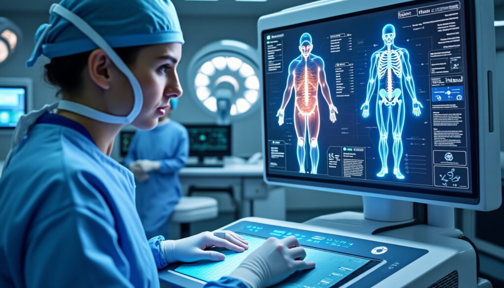
Key technological milestones include 3D functional MRI systems, which provide detailed neuron activity maps crucial to spinal surgery and neuro-oncology. Innovations like intraoperative MRI facilitate immediate assessment of surgical impact, allowing real-time corrective measures and ensuring thorough lesion resection. In parallel, fluorescence imaging advances, particularly near-infrared technology, support precise tissue differentiation in complex oncological procedures.
Another major breakthrough lies in extended reality (XR) and artificial intelligence integration. XR platforms overlay medical imaging data onto the surgeon’s field of view, bolstering spatial awareness and surgical planning accuracy. AI algorithms assist in image interpretation, anomaly detection, and outcome prediction, effectively augmenting clinical decision making. This combination accelerates the surgical workflow and reduces cognitive load, which is vital during intricate spinal operations.
Additionally, robotic-assisted platforms tied to imaging diagnostics have expanded capabilities to perform minimally invasive spinal surgeries with greater dexterity. The FDA-approved eCential Robotics system exemplifies this evolution, combining 3D imaging and robotic navigation to minimize human error and optimize implant positioning.
These technological advances collectively highlight a shift from reactive to proactive surgical management, positioning imaging as both a diagnostic and therapeutic cornerstone. The future of surgery indeed hinges on the continuous integration of innovative imaging solutions, amplifying surgeons’ capabilities and substantially improving patient outcomes.
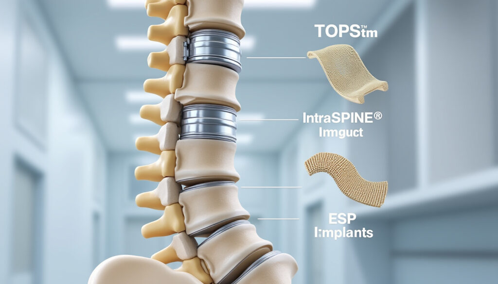
The evolving field of spinal surgery is witnessing a transformative shift, fueled by cutting-edge implant technologies such as TOPS, Intraspine, and ESP. These innovative implants redefine treatment possibilities by emphasizing preservation of natural spinal mechanics, while ensuring durable stabilization and…
Organizing Imaging Exams: Best Practices for Surgical Precision and Safety
Organizing imaging exams systematically is fundamental for delivering high-quality surgical care. Best practices emphasize structured protocols, personalized imaging pathways, and multidisciplinary coordination to ensure accurate diagnostics and optimized preparation for surgery. Applying these practices enhances surgical precision and reduces the risk of complications.
Core principles include:
- Timely scheduling: Avoiding delays between imaging acquisition and surgical intervention ensures relevance and accuracy of diagnostic data.
- Protocol standardization: Tailoring imaging techniques and sequences to specific pathologies and surgical needs enhances image quality and interpretability.
- Multidisciplinary engagement: Collaboration between radiologists, surgeons, and ancillary staff fosters consensus on imaging strategy and interpretation.
- Data integration: Utilizing centralized, accessible imaging databases encourages streamlined information flow and facilitates intraoperative utilization.
- Patient-centered approach: Providing clear explanations and education reduces anxiety and empowers patients in informed consent processes.
Adopting these practices leads to measurable improvements in surgical outcomes by permitting refined preoperative visualization and anticipating intraoperative challenges. Moreover, enhanced imaging organization supports personalized postoperative care by providing baseline imaging for recovery monitoring and early complication detection.
The dynamic nature of surgical innovations requires that imaging exam management remain adaptable and responsive. The ongoing education of healthcare teams on new imaging modalities and their clinical application ensures sustained excellence in surgical precision and patient safety.

Preparing for Surgery in France: Key Indications, Benefits, and Limitations
In an evolving global healthcare landscape, France emerges as a beacon of advanced surgical expertise, particularly attracting international patients seeking expedited and innovative medical interventions. Patients from regions experiencing long wait times, such as Quebec, increasingly view France as an…
Comparative Overview of Innovative Imaging Technologies Empowering Spine Surgery
Modern spine surgery benefits from a spectrum of advanced imaging technologies, each contributing distinct advantages to surgical planning and execution. The table below highlights key modalities, functionalities, and their impacts on clinical outcomes, illustrating how strategic imaging management aligns with surgical innovation.
| Imaging Technology | Primary Function | Advantages in Surgery | Example Application |
|---|---|---|---|
| 3D Functional MRI | Mapping neuronal activity | Preserves neurological function; guides preoperative planning for tumors and myelopathies | Planning surgeries for complex spinal cord tumors |
| Computer-Assisted Navigation | Real-time anatomical visualization | Enhances millimeter precision; reduces invasive incisions | Minimally invasive spinal fusion procedures |
| Near-Infrared Fluorescence Imaging | Visualizes lymphatic and tissue structures | Reduces post-op complications; improves oncological surgery safety | Lymphatic mapping in spinal oncology |
| Robotic-Assisted Surgery Platforms | Augments surgeon dexterity and control | Increases precision; shortens operative times; facilitates complex procedures | Robotic-guided spinal instrumentation placement |
This comparative insight underscores the need for comprehensive imaging exam coordination to fully harness these technologies. Ensuring compatibility and real-time accessibility is essential to embed these innovations into everyday surgical practice effectively.
The Future of Surgery Hinges on the Strategic Management of Imaging Exams
Explore the cutting-edge medical imaging technologies that revolutionize spine surgery by enhancing precision and patient outcomes.
As you type, the list of technologies below will update to match your keyword.

Unveiling the Hidden Benefits of Quality and Certification in Devices
The landscape of device manufacturing and clinical application in 2026 has been fundamentally shaped by the rigorous interplay between quality management and certification processes. Within this complex framework, the hidden benefits of adhering to international standards and obtaining relevant certifications…
Enhancing Clinical Decision Making through Structured Imaging Exam Protocols
Structured imaging exam protocols directly empower clinical decision making by delivering consistent, high-quality data focused on the pathological and anatomical subtleties inherent to each patient’s condition. Protocol standardization is becoming a cornerstone to reducing errors, ensuring reproducibility, and improving the comparative analysis of sequential imaging.
For surgical candidates, this approach enables selective imaging aligned with the clinical question—avoiding excessive testing or irrelevant data acquisition. Imaging sequences customized for specific spine pathologies, such as herniated discs or cervical myelopathies, provide surgeons with crisp images delineating nerve root compression or ischemic changes. This precision informs the choice of conservative management versus surgical intervention and the tailoring of surgery type accordingly.
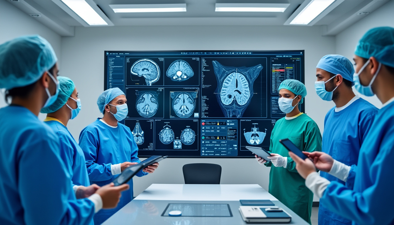
Furthermore, structured protocols facilitate interdisciplinary communication by ensuring that radiologists and surgeons interpret images through a shared understanding of terminology, quality control, and clinical intent. This reduces ambiguities that might arise in uncoordinated imaging practices, strengthening collaborative planning and patient counseling.
For patients, such clarity in imaging and decision making fosters trust and adherence to treatment plans. It also complements post-surgical follow-up by enabling consistent comparability in longitudinal imaging studies, crucial to detecting complications or disease progression.
Current literature increasingly advocates for the widespread adoption of structured imaging exam protocols as an ethical and quality imperative in surgical care. This aligns with advances in artificial intelligence, which depend on standardized imaging inputs for optimal algorithmic support, further amplifying clinical decision making capabilities.
Why Experts Advocate for Comprehensive Preoperative Evaluations
In the complex and demanding field of spinal surgery, the emphasis on comprehensive preoperative evaluations has become a fundamental pillar supporting successful surgical outcomes. These evaluations, far beyond mere routine checks, integrate detailed analyses of patient health, surgical risk assessments,…
Personalized Patient Care Through Coordinated International Imaging and Surgical Pathways
Patient outcomes reach their highest potential when surgical innovation is paired with personalized, coordinated care pathways. Strategic management of imaging exams plays a pivotal role in this synthesis, especially when patients undergo complex surgeries abroad, requiring seamless international coordination.
The collaborative ecosystem engaging neurotherapeutic specialists, surgical teams, and medical logistics entities exemplifies this model. Patients from Quebec planning spinal surgery in France experience a tailored journey beginning with comprehensive neurofunctional evaluations and advanced imaging analyses at home. Coordination teams oversee the precise scheduling and transfer of imaging data while aligning clinical goals between continents.
This detailed preparation facilitates informed surgical planning featuring state-of-the-art implants and minimally invasive approaches, such as dynamic stabilization devices like TOPS. The system extends beyond the operating room, integrating postoperative rehabilitation programs that utilize biofeedback and neurofeedback derived from continuous imaging monitoring.
Such personalized pathways reduce surgical risks by identifying contraindications early, anticipating complications, and optimizing biomechanical restoration. They also help patients navigate administrative and logistical challenges, reducing stress and promoting adherence to rehabilitation protocols essential for long-term recovery.
This integrative approach represents the epitome of how the future of surgery hinges on the strategic management of imaging exams combined with human-centered coordination. It reassures patients that cutting-edge technology and compassionate care converge for optimal outcomes.
The Future of Surgery Lies in Stepwise Functional Recovery
The evolution of surgical care in 2026 reveals a compelling shift toward approaches that prioritize the holistic recovery of patients, moving beyond traditional metrics like length of hospital stay or mortality rates. This modern paradigm highlights the principle that the…
Identifying Pathologies and Surgical Techniques Enabled by Advanced Imaging Coordination
Advanced imaging coordination expands the array of spinal pathologies that can be addressed more effectively through surgical innovation. Detailed diagnostic images feed directly into customized surgical plans that incorporate the latest minimally invasive and motion-preserving techniques.
Examples of targeted conditions and corresponding surgical solutions include:
- Herniated lumbar and cervical discs: Treated via microdiscectomy or disc replacement prostheses to restore segmental mobility.
- Spinal canal stenosis: Managed with minimally invasive decompression and, if necessary, dynamic stabilization implants.
- Spondylolisthesis: Corrected through spinal fusion or dynamic implants prioritizing mobility preservation.
- Adult scoliosis and spinal deformities: Addressed with corrective surgery employing advanced implants like the TOPS system for dynamic support.
- Cervical myelopathies: Treated with image-guided decompression to prevent further neurological deterioration.
Technologies facilitating these surgeries, such as robotic navigation systems and fluorescence imaging, rely on well-managed imaging exam workflows. Without precise, accessible imaging data, many of these sophisticated interventions would risk higher complication rates or suboptimal outcomes.
This integration ensures that each patient receives a treatment plan tailored to their unique pathology and anatomical considerations, validated by cutting-edge imaging diagnostics. International networks further broaden access to these innovations, enhancing global standards of spinal care.
{“@context”:”https://schema.org”,”@type”:”FAQPage”,”mainEntity”:[{“@type”:”Question”,”name”:”How can I determine if I am a suitable candidate for spine surgery abroad?”,”acceptedAnswer”:{“@type”:”Answer”,”text”:”An in-depth neurofunctional evaluation combined with a comprehensive review of imaging exams and clinical questionnaires guides eligibility. Experts in Quebec coordinate this process, ensuring appropriate surgical recommendations based on individual pathology.”}},{“@type”:”Question”,”name”:”What imaging documents are required for preoperative assessment?”,”acceptedAnswer”:{“@type”:”Answer”,”text”:”Complete sets of prior imaging studies such as MRI, CT, and X-rays, along with relevant medical records, are necessary. Detailed clinical questionnaires complement these, facilitating precise evaluation and fostering effective collaboration with surgical centers abroad.”}},{“@type”:”Question”,”name”:”What happens after surgery performed in an international facility?”,”acceptedAnswer”:{“@type”:”Answer”,”text”:”Postoperative care is coordinated by specialist teams who schedule follow-up appointments, personalize rehabilitation protocols involving biofeedback and neurofeedback, and provide continuous support to ensure safe recovery and minimize complications.”}},{“@type”:”Question”,”name”:”How does international coordination improve my surgical journey?”,”acceptedAnswer”:{“@type”:”Answer”,”text”:”Integrated partnerships among neurotherapy clinics, medical logistics, specialized surgical teams, and patient support organizations reduce wait times, streamline administrative procedures, and ensure seamless clinical continuity, enhancing overall care quality.”}},{“@type”:”Question”,”name”:”What surgical technologies offered abroad are superior to local options?”,”acceptedAnswer”:{“@type”:”Answer”,”text”:”Access to advanced implants like TOPS and Intraspine, minimally invasive techniques, and cutting-edge image-guided surgical planning enables faster recovery and preservation of spinal mobility compared to conventional systems.”}}]}How can I determine if I am a suitable candidate for spine surgery abroad?
An in-depth neurofunctional evaluation combined with a comprehensive review of imaging exams and clinical questionnaires guides eligibility. Experts in Quebec coordinate this process, ensuring appropriate surgical recommendations based on individual pathology.
What imaging documents are required for preoperative assessment?
Complete sets of prior imaging studies such as MRI, CT, and X-rays, along with relevant medical records, are necessary. Detailed clinical questionnaires complement these, facilitating precise evaluation and fostering effective collaboration with surgical centers abroad.
What happens after surgery performed in an international facility?
Postoperative care is coordinated by specialist teams who schedule follow-up appointments, personalize rehabilitation protocols involving biofeedback and neurofeedback, and provide continuous support to ensure safe recovery and minimize complications.
How does international coordination improve my surgical journey?
Integrated partnerships among neurotherapy clinics, medical logistics, specialized surgical teams, and patient support organizations reduce wait times, streamline administrative procedures, and ensure seamless clinical continuity, enhancing overall care quality.
What surgical technologies offered abroad are superior to local options?
Access to advanced implants like TOPS and Intraspine, minimally invasive techniques, and cutting-edge image-guided surgical planning enables faster recovery and preservation of spinal mobility compared to conventional systems.